Discovery in zebrafish may yield better understanding of a range of mental health disorders related to stress, anxiety
Ma et al., 2020b
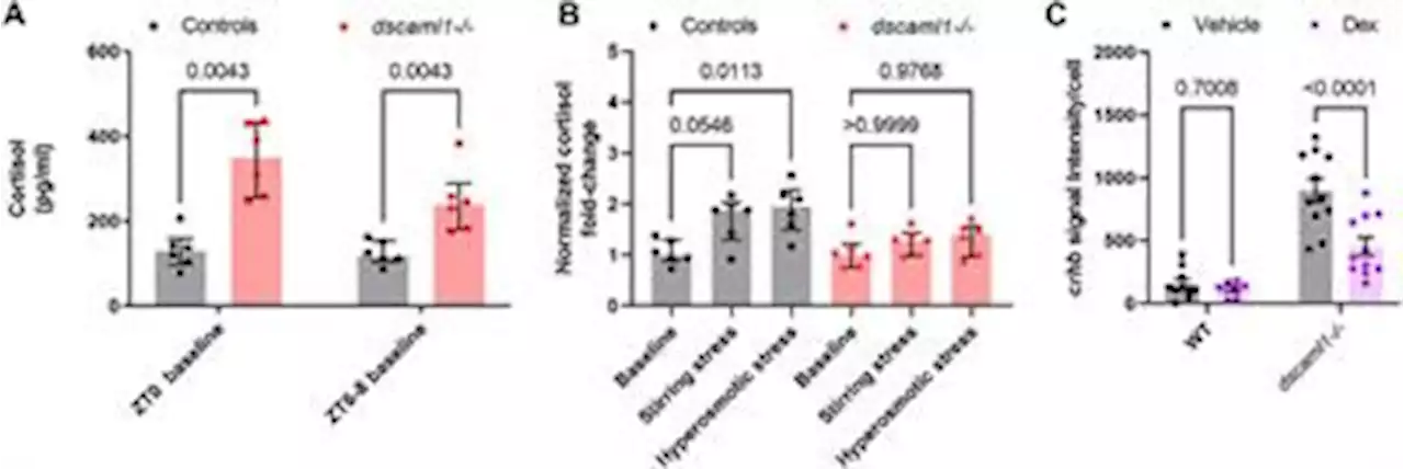
). 30–35 animals were placed in each petri dish for cortisol extraction. The morning baseline sample collections were done at 15–30 min after light onset , and the afternoon sample collections were done between 14:30–16:00. Hyperosmotic stress and stirring stress experiments were done between 14:30–15:30. 6 biological duplicates—each containing a pool of 30 animals—were collected for each genotype and stress condition . Mutants and control animals are tested side by side for each experiment.
Stirring stress was induced by creating a vortex water flow with a spinning magnetic stir bar . A small magnetic stir bar was placed into a 100 mm petri dish containing 35 animals and 20 mL of E3 media. The stir bar was rotated at 300 rpm with a stirring microplate for 5 min. Hyperosmotic stress was induced by increasing salt concentration in the media . 30 animals were placed in 8 mL of E3 media. Then, 2 mL of prewarmed 1.
Sample homogenization and cortisol extraction were performed as described by Yeh et al. . Briefly, 5 dpf larvae were rapidly immobilized with ice-cold E3 media and then flash-frozen at −80°C. Once all samples were collected, cortisol from the frozen samples was extracted with ethyl acetate . Cortisol concentration was measured using a commercial ELISA kit, following the manufacturer’s instructions . Sample plates were read with a microplate reader 90–120 min after initial development.
Two-photon live imaging was done on a custom Bruker two-channel two-photon microscope. A tuneable Ti:Saphire laser was tuned to 980 nm to excite RFP and GFP simultaneously. 78 hpf animals were anesthetized and embedded the same way as confocal live imaging, but with the dorsal side away from the cover glass. Each animal was imaged at 78, 84, 96, 108, and 120 hpf . After each time point, imaged larvae were gently removed from the agarose and recovered in E3 media at 28.
Belgique Dernières Nouvelles, Belgique Actualités
Similar News:Vous pouvez également lire des articles d'actualité similaires à celui-ci que nous avons collectés auprès d'autres sources d'information.
 Frontiers | Neuroplasticity in F16 fighter jet pilotsExposure to altered g-levels causes unusual sensorimotor demands that must be dealt with by the brain. This study aimed to investigate whether fighter pilots, who are exposed to frequent g-level transitions and high g-levels, show differential functional characteristics compared to matched controls, indicative of neuroplasticity. We acquired resting-state functional magnetic resonance imaging data to assess brain functional connectivity (FC) changes with increasing flight experience in pilots and to assess differences in FC between pilots and controls. We performed whole-brain exploratory and region-of-interest (ROI) analyses, with the right parietal operculum 2 (OP2) and the right angular gyrus (AG) as ROIs. Our results show positive correlations with flight experience in the left inferior and right middle frontal gyri, and in the right temporal pole. Negative correlations were observed in primary sensorimotor regions. We found decreased whole-brain FC of the left inferior frontal gyrus in fighter pilots compared to controls and this cluster showed decreased FC with the medial superior frontal gyrus. FC increased between the right OP2 and the left visual cortex, and between the right left AG in pilots compared to controls. These findings suggest altered motor, vestibular, and multisensory processing in the brains of fighter pilots, possibly reflecting coping strategies to altered sensorimotor demands during flight. Altered FC in frontal areas may reflect adaptive cognitive strategies to cope with challenging conditions during flight. These findings provide novel insights into brain functional characteristics of fighter pilots, which may be of interest to humans traveling to space.
Lire la suite »
Frontiers | Neuroplasticity in F16 fighter jet pilotsExposure to altered g-levels causes unusual sensorimotor demands that must be dealt with by the brain. This study aimed to investigate whether fighter pilots, who are exposed to frequent g-level transitions and high g-levels, show differential functional characteristics compared to matched controls, indicative of neuroplasticity. We acquired resting-state functional magnetic resonance imaging data to assess brain functional connectivity (FC) changes with increasing flight experience in pilots and to assess differences in FC between pilots and controls. We performed whole-brain exploratory and region-of-interest (ROI) analyses, with the right parietal operculum 2 (OP2) and the right angular gyrus (AG) as ROIs. Our results show positive correlations with flight experience in the left inferior and right middle frontal gyri, and in the right temporal pole. Negative correlations were observed in primary sensorimotor regions. We found decreased whole-brain FC of the left inferior frontal gyrus in fighter pilots compared to controls and this cluster showed decreased FC with the medial superior frontal gyrus. FC increased between the right OP2 and the left visual cortex, and between the right left AG in pilots compared to controls. These findings suggest altered motor, vestibular, and multisensory processing in the brains of fighter pilots, possibly reflecting coping strategies to altered sensorimotor demands during flight. Altered FC in frontal areas may reflect adaptive cognitive strategies to cope with challenging conditions during flight. These findings provide novel insights into brain functional characteristics of fighter pilots, which may be of interest to humans traveling to space.
Lire la suite »
 Plans to change function room into homes could be pub's lifelineThe first floor of the pub could be converted if the plans are passed by councillors
Lire la suite »